Treatments


How can dry AMD be treated?
The most effective treatment for dry AMD is a vitamin/mineral combination as outlined in the Age Related Eye Disease Study, AREDS. This is the only proven regimen. This government sponsored study, involving more than 4500 people over more than 10 years found that a treatment with Vitamin A, C, E, Zinc and Copper can significantly decrease the risk for vision loss if taken at the right dosage and when "enough" disease has developed. The supplements do not seem to prevent the development of AMD itself. At the same time a follow up study, AREDS 2, is currently investigating if Lutein-like drugs and omega 3 fish-oil have a similar protective effect. None of this will cure AMD but may help delay or avoid vision loss. Also, simply having a family history of AMD is not sufficient reason to start AREDS vitamins as the vitamins do not prevent the onset of AMD. Please ask our doctors which treatment may be best for you.
The AREDS vitamins formula is as follows:
- Vitamin A: 15 mg equivalent to 25,000 IU
- Vitamin C: 500 mg
- Vitamin E: 400 IU
- Zinc: 80 mg
- Copper: 2 mg
How can wet AMD be treated?
Wet macular degeneration can now very often be halted and in some cases even be reversed to some degree with the help of drugs that are injected into the eye. These drugs, known as anti-VEGF are scientific miracles having helped millions of patients stabilize and improve their vision for a disease that until recently had limited treatments and lead to serious changes in one's life.
Researchers have found that a chemical called vascular endothelial growth factor, or VEGF, is critical in causing abnormal blood vessels to grow under the retina. Scientists have developed several new drugs that can block the trouble-causing VEGF. These are referred to as anti-VEGF drugs, and they help block abnormal blood vessels, slow their leakage, and help reduce vision loss.
Treatment with the anti-VEGF drug is usually performed by injecting the medicine with a very fine needle into the back of your eye. Prior to that the eye is cleaned and anesthetic drops or medicine is administered to reduce eye pain. Usually, patients receive multiple anti-VEGF injections over the course of many months. There is a small risk of complications with anti-VEGF treatment, usually resulting from the injection itself. However, for most people, the benefits of this treatment outweigh the small risk of complications.
Anti-VEGF medications are a step forward in the treatment of wet AMD because they target the underlying cause of abnormal blood vessel growth. This treatment offers new hope to those affected with wet AMD. Although not every patient benefits from anti-VEGF treatment, a large majority of patients achieve stabilized vision, and a significant percentage can improve to some degree. We have been treating our patients with Avastin, Lucentis, and more recently Eylea.
In some cases, the injections can be combined with the special laser treatments.
What is the future of AMD treatments?
AMD is one of the most extensively studied and researched areas in ophthalmology. We are learning more about the genes implicated in the disease as well as new treatments. There are multiple therapies that being studied in clinical practices and academic institutions throughout the world. There is promising research on going to find ways to slow down dry AMD. Also, ways to extend the treatment for wet AMD patients is being evaluated with different treatment protocols and new therapies. The future of patients suffering with AMD is promising as it has ever been with newer treatment options will likely be available over the next several years.
For information and consultation regarding treatment for Macular Degeneration, contact our main office at (405) 948-2020.
There are several treatments for diabetic retinopathy: laser surgery, intraocular injections of medications to reduce retinal swelling or to suppress blood vessel growth, and vitrectomy. Combinations of these treatments are very effective in reducing vision loss from this disease. In fact, even people with advanced retinopathy have an excellent chance of keeping their vision when they are treated early. However, it is important to note that although these treatments are very successful, they do not cure diabetic retinopathy, and they also do not cure your underlying diabetes. Therefore proper control of blood sugar, blood pressure, and cholesterol is important.
All treatments for diabetic eye disease involve cooperation with your family doctor, internist or endocrinologist as very often diabetes eye disease is a sign that the underlying medical conditions may need closer and more aggressive monitoring.
If the retina is detached, it must be reattached before sealing the retinal tear. There are three ways to repair retinal detachments. Pneumatic retinopexy involves injecting a special gas bubble into the eye that pushes on the retina to seal the tear. This procedure is typically performed in our office. The scleral buckle procedure requires the fluid to be drained from under the retina before a flexible piece of silicone is sewn on the outer eye wall to give support to the tear while it heals. Most often retinal detachment repair involves Vitrectomy surgery which removes the vitreous gel from the eye, replacing it with a gas bubble, which is slowly replaced by the body fluids. Laser treatment or cryotherapy (freezing) treatment is applied at the time of surgery to treat the areas of torn retina.
Vitrectomy is a type of eye surgery used to treat disorders of the retina (the light-sensing cells at the back of the eye) and vitreous (the clear gel-like substance inside the eye). It may be used to treat a severe eye injury, diabetic retinopathy, retinal detachments, macular pucker (wrinkling of the retina), and macular holes.
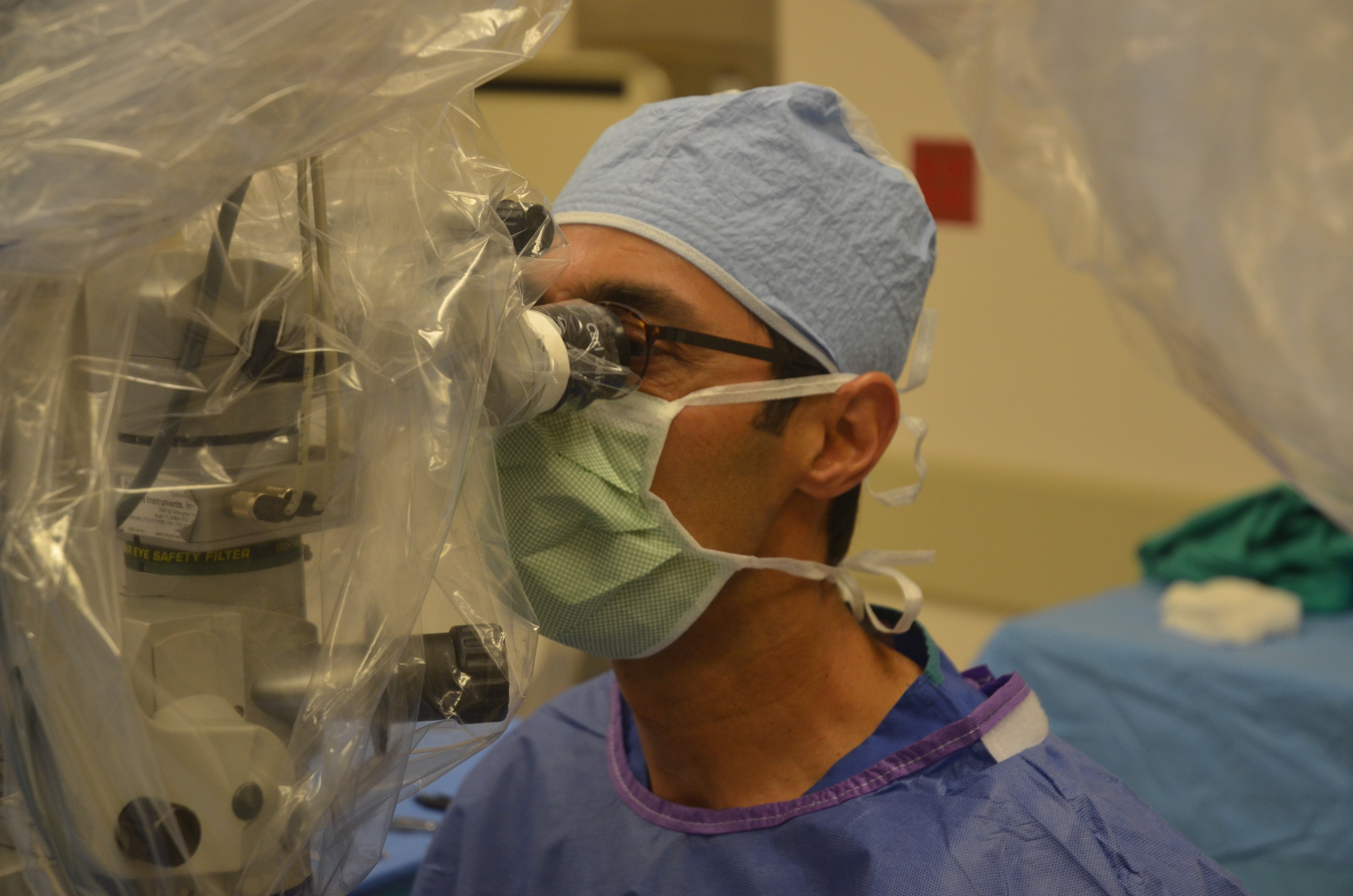
During a vitrectomy operation, the surgeon makes tiny incisions in the sclera (the white part of the eye). Using a microscope to look inside the eye and microsurgical instruments, the surgeon removes the vitreous and repairs the retina through these tiny incisions. Repairs include removing scar tissue or a foreign object if present.
During the procedure, the retina may be treated with a laser to reduce future bleeding or to fix a tear in the retina. An air or gas bubble that slowly disappears on its own may be placed in the eye to help the retina remain in its proper position, or a special fluid that is later removed may be injected into the vitreous cavity.
Oklahoma Retinal Consultants performs vitrectomy only in an operating room setting with the expertise of an anesthesiologist. Anesthesia for the procedure typically is sedation with a local anesthetic around the eye.
Recovering from vitrectomy surgery may be uncomfortable, but the procedure often improves or stabilizes vision. Once the blood- or debris-clouded vitreous is removed and replaced with a clear medium (often a saltwater solution), light rays can once again focus on the retina. Vision after surgery depends on how damaged the retina was before surgery.
Vitrectomy surgery, the only treatment for a macular hole, removes the vitreous gel and scar tissue pulling on the macula and keeping the hole open. The eye is then filled with a special gas bubble to push against the macula and close the hole. The gas bubble will gradually dissolve, but the patient must maintain a face-down position for five days to keep the gas bubble in contact with the macula. Success of the surgery often depends on how well the position is maintained. Our macular hole repair rate with single surgery is 98%. Macular holes can close with upright posture but our recommendation is face down positioning for a few days at a minimum if possible to maximize success rate.
With treatment, most macular holes shrink, and some or most of the lost central vision can slowly return. The amount of visual improvement typically depends on the length of time the hole was present and damage that may have already occurred.
Laser treatment is used to treat multiple types of retinal disorders. A laser by definition allows a focused beam of light at a specific wavelength. Inside the eye this can be used to treat retinal tears, leaking or new blood vessels, lack of blood flow, or prevent small detachments from worsening. The effect of the laser is mostly from the scar it produces in the retina and choroid. Laser treatment usually leads to minimal discomfort but when extended treatment is required, some individuals can experience discomfort which can require additional local anesthesia. Laser treatment can be administered both in the clinic and operating room.
Cryotherapy involves the use of a probe that touches the white part of eye (sclera) which produces a tissue reaction that leads to formation of a controlled retinal scar. This is helpful in patients undergoing repair of retinal tear or detachment. Treatment can be administered both in the office and operating room as necessary. As cryotherapy can sometimes be painful, Our doctors utilizes local anesthetic agent prior to its use and patients are asked to use pain relievers for the first day.
An intravitreal injection allows administering a drug or substance inside the eye (vitreous cavity). This allows the highest concentration of medication to reach the inside the eye and is utilized in patients suffering age related macular degeneration and diabetic eye disease. This technique is also utilized with injection of a small gas bubble in the eye for in office treatment of limited retinal detachment.
This is an outpatient procedure involving the use of a special light-activated drug is used to treat some patients with wet AMD. PDT causes fewer visual side effects than other treatments. The benefit of PDT is that it inhibits abnormal blood vessel leakage associated with wet macular degeneration, limiting damage to the overlying retina.
With PDT, the inactive form of the drug is usually injected into a vein in the arm, where it travels to and accumulates in abnormal blood vessels under the center of the macula. A special low-intensity laser light targeted at the retina activates the drug only in the affected area, damaging the abnormal blood vessels under the retina and leaving normal blood vessels intact.
Patients who are treated with PDT will become temporarily extra sensitive to bright light (photosensitive). Care should be taken to avoid exposure of the skin or eyes to direct sunlight or bright indoor light for several days.
PDT therapy is not effective for treatment of atrophic or AMD, which is caused by aging and thinning of the tissues of the macula. Although photodynamic therapy can preserve vision for many people, it may not stop vision loss in all patients. The abnormal blood vessels may regrow or begin to leak again. PDT treatments are often combined with intravitreal steroid or anti-VEGF treatments. Multiple PDT treatments sometimes are necessary.

